Patients frequently ask: ‘If I have an ultrasound, can it see the cervical cancer?’
The answer is yes, but with an important distinction between detection and screening.
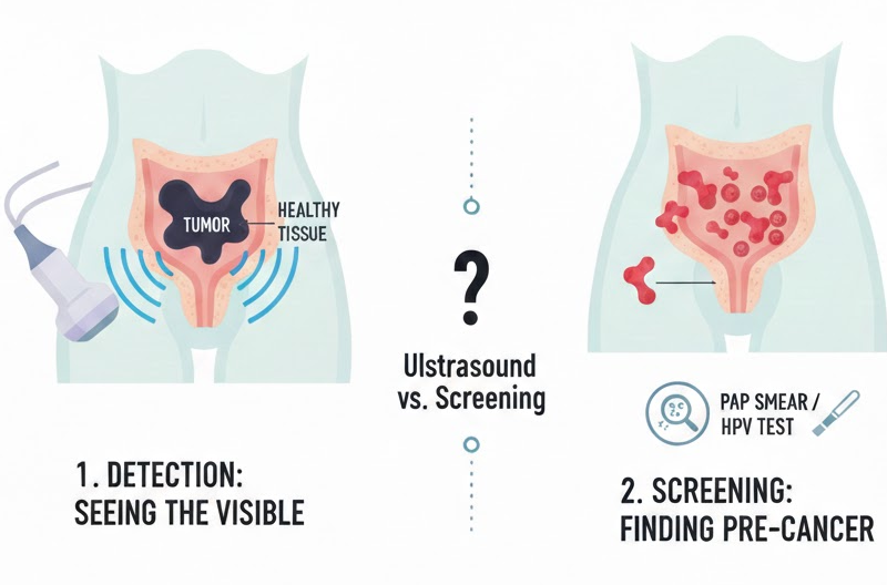
1. Detection: Seeing the Visible Tumor
Modern high-resolution ultrasound, specifically Transvaginal (TVS) and Transrectal (TRS), is a powerful tool for detecting and visualizing cervical tumors. When a tumor has reached a certain size, it becomes clearly visible to the “eye” of the ultrasound.
- High Sensitivity: Clinical studies show that in women with suspected or confirmed cervical cancer, ultrasound has a sensitivity of approximately 80% to 93% for identifying the tumor. This makes it an incredibly reliable tool for initial radiological assessment.
- On an ultrasound scan, cervical cancer typically appears as a hypoechoic mass. This means the tumor appears as a darker, denser area compared to the bright, uniform, and healthy tissue of the surrounding cervix.
2. Why Ultrasound is Not a Screening Tool
If ultrasound is so effective at “seeing” the cancer, why don’t we use it instead of a Pap smear or HPV test for yearly checkups? The reason lies in the nature of early-stage disease:
- Missing “Pre-Cancer”: Screening is designed to find microscopic cellular changes (dysplasia) years before they ever turn into a physical tumor. Ultrasound cannot “see” individual cells; it only sees a mass once it has already formed.
- The “Hidden” Phase: In the very earliest stages, a small cancer may be flush with normal tissue or tucked inside the cervical canal, making it invisible to sound waves.
- The Need for Confirmation: While ultrasound can identify a “suspicious growth,” it cannot provide a definitive diagnosis. Only a biopsy (analyzing a tissue sample under a microscope) can confirm if a mass is malignant.
3. Beyond Detection: The “Superpower” of Management
3. Beyond Detection: Ultrasound and Management
Once a diagnosis is confirmed via biopsy, ultrasound shifts from being a detection tool to being a management powerhouse. This is where your radiology team provides the most value to your treatment plan.
We use ultrasound to “map” the cancer in ways that a physical exam cannot:
- Measuring the “Roots”: We assess the depth of stromal invasion—essentially seeing how deep the cancer goes into the cervical wall.
- Checking the Neighbors: We evaluate if the mass is touching or invading nearby structures like the bladder, the rectum, or the parametrium (the tissue next to the uterus).
- Surgical Planning: For patients hoping to preserve their fertility, ultrasound provides the precise measurements needed to determine if fertility-sparing surgery is a safe option.
If it has been a while since your last check-up, an ultrasound is an excellent place to start; it provides an immediate look to ensure no visible masses or tumors are present. However, because early cellular changes are invisible to the naked eye, we always recommend following your scan with a Pap smear or HPV test. Together, these tools offer the ultimate reassurance: the ultrasound confirms you are clear today, and screening ensures you stay protected for tomorrow.”
Contact us to book your ultrasound!

No comment